info@chuka.ac.ke
020 231 0512/ 020 231 0518
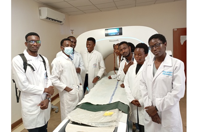
The students began their tour at the CT Scan Unit, where they learned about head CT scans and the use of contrast agents to enhance diagnostic accuracy by visualizing blood vessels. This introduction underscored the importance of precision in diagnosing complex medical conditions.
Next, the group visited the Digital Radiography (X-Ray) department, where they observed the advantages of digital X-rays over traditional methods. They learned how these technologies improve image quality, reduce radiation exposure, and streamline the diagnostic process, providing faster and safer results for patients.
At the Dental X-Ray Center, the students gained insights into the role of dental imaging in identifying cavities, infections, and other oral health issues. This session highlighted the significance of specialized imaging in preventive and restorative dental care.
The Mammography Unit showcased the critical role of imaging in early breast cancer detection. Radiographers explained the life-saving potential of mammography in identifying abnormalities at stages when treatment is most effective. The visit also included the Ultrasound Facility, where students explored how ultrasound imaging is used to diagnose conditions in soft tissues and organs, as well as to monitor pregnancies.
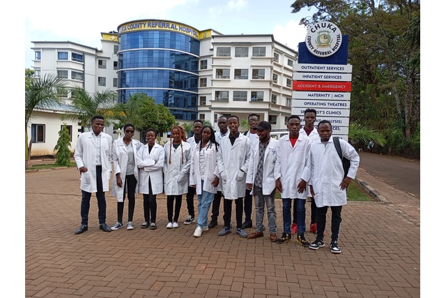
The students were actively engaged throughout the visit, asking questions and taking detailed notes as the hospital’s radiographers shared their expertise. The experience offered a comprehensive understanding of the pivotal role that medical imaging plays in diagnosing and managing a wide range of health conditions.
Chuka University is …… Inspiring Environmental Sustainability for Better Life